
A young footballer rolls his ankle in a tackle. He can walk on it, so he is told it is just a sprain. Two weeks later the pain still has not settled, so his GP orders an MRI. The report comes back: syndesmosis intact. Reassured, he returns to training and but never quite gets back to pre-injury level.
This scenario plays out in my rooms regularly. New research from our team at Melbourne Orthopaedic Group, published this month in Foot and Ankle Surgery, suggests it may be happening more often than anyone realised, not because MRI itself is a poor test, but because the written report frequently does not reflect what is actually on the scan.
Syndesmosis injuries, also known as high ankle sprains, involve the ligaments joining the shin bone (tibia) and the outer lower leg bone (fibula) just above the ankle joint. Unlike a common rolled ankle, they are notoriously easy to miss on first assessment, and an MRI can be the decisive investigation that determines whether a patient is managed conservatively or referred for surgical stabilisation. Our new paper asks a simple but important question: how much can we trust the words in that MRI report?
Why We Looked at This
I see patients most weeks who arrive with an MRI report stating the syndesmosis, the group of ligaments joining the tibia and fibula just above the ankle, is intact or unremarkable, yet who go on to test unstable on clinical examination or at arthroscopy. Referring clinicians, understandably, rely on the written report rather than reviewing the images themselves, because that is what reaches their desk. If that report under-calls the injury, the diagnosis is delayed and so is the correct treatment.
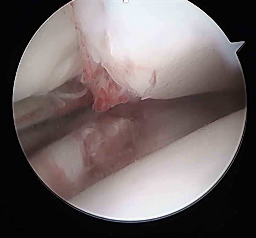
An unstable syndesmosis as viewed during ankle arthroscopy (drive through sign).
Together with our team Edina Vacariu, Alessia Tevere, David Shepherd and Tim Schneider, I set out to test this formally. We reviewed 73 patients who underwent both an MRI and an ankle arthroscopy at our institution between August 2024 and September 2025. Only MRI reports that specifically commented on the syndesmosis were included. We compared what the radiology report said about the syndesmosis against what we actually found at arthroscopy, which is the gold standard for diagnosing syndesmotic instability, using the drive-through test described by Guyton. The cohort included 28 women and 45 men, average age 36, with a roughly even split between acute injuries (36 patients) and chronic injuries present for more than six weeks (37 patients). Nine patients also had an associated fracture.
What We Found
The results were striking.
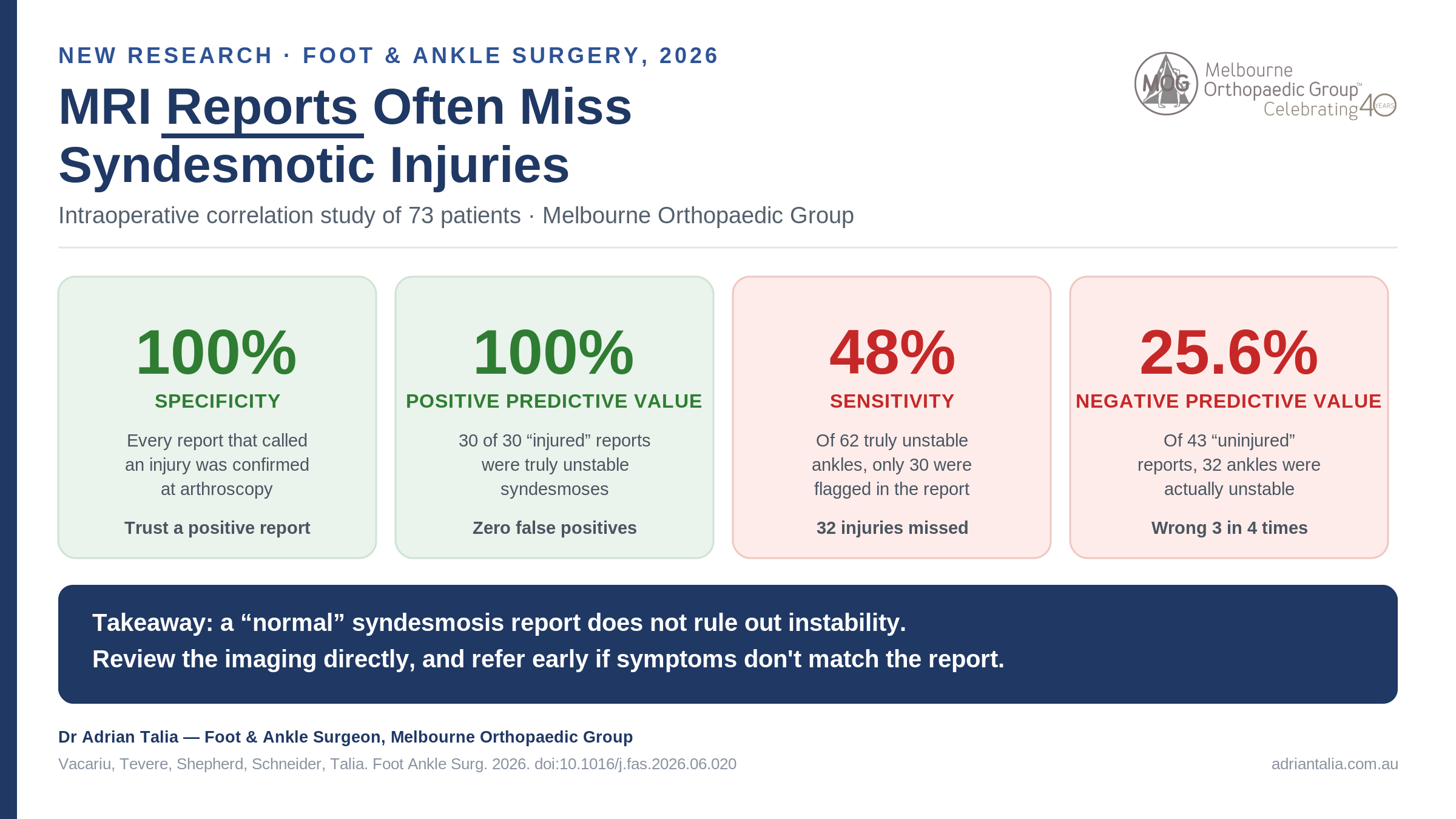
When an MRI report described the syndesmosis as injured, it was right every time. All 30 of these cases were confirmed unstable at arthroscopy, giving a specificity and positive predictive value of 100%. In plain terms, if the report says the syndesmosis is torn, you can believe it.

MRI and weight bearing CT imaging of an unstable syndesmosis.
The problem sits on the other side. Of the 43 reports that described the syndesmosis as intact or uninjured, only 11 were genuinely stable at arthroscopy. The remaining 32 were unstable, missed entirely by the written report. That works out to a sensitivity of just 48% and an overall accuracy of 56.2%. A report stating “uninjured syndesmosis” turned out to be wrong roughly three times out of four when the ankle was in fact unstable, a negative predictive value of only 25.6%.
Think of it like a smoke alarm that never gives a false alarm but regularly fails to sound when there really is a fire. That is what our data showed: a test that is trustworthy when positive, but one you cannot rely on to reassure you when it comes back negative.
Why the Gap Exists
This is not a criticism of MRI as a technology. When ankle images are read using defined diagnostic criteria by someone experienced in musculoskeletal imaging, MRI performs extremely well, with sensitivity and specificity both above 85% in the published literature. Our study measured something different: how well the final, dictated report, the document a GP or physiotherapist actually receives and acts on, reflects the underlying pathology.
In our health system, ankle MRIs are ordered by a wide range of clinicians and read by a wide range of radiologists, not all of whom have subspecialty musculoskeletal training. There is also no standardised reporting template for the ankle syndesmosis, so two radiologists looking at similar images may describe them quite differently. The imaging data may well contain everything needed for an accurate diagnosis. It is the interpretation and distillation of that data into a written report where the accuracy is being lost.
Interestingly, when a patient in our cohort also had a fibula fracture, MRI reporting accuracy was 100%. A visible fracture is an obvious prompt to look carefully at the syndesmosis. It is the isolated ligamentous injury, the subtle, easily overlooked high ankle sprain without a broken bone, that the written report is most likely to miss.
What This Means for Diagnosis and Referral
This has real consequences. We know from previous research that delayed treatment of an unstable syndesmosis leads to worse functional outcomes, and that outcomes are best when stabilisation surgery happens within six weeks of injury. If a GP or physiotherapist reads “syndesmosis intact” and closes the loop on referral, a genuinely unstable ankle can go untreated for months, setting up chronic instability and early degenerative change in the joint.
My advice, and the conclusion of our paper, is straightforward. A report stating the syndesmosis is uninjured should be viewed with some caution, particularly in a patient whose symptoms are not settling as expected. Persistent pain above the ankle joint line, difficulty pushing off, or a sense of instability after a “normal” MRI report still warrants a specialist opinion. I always review the MRI images myself rather than relying solely on the written report, and I correlate this with the history and clinical examination: the squeeze test, the external rotation test, and where needed, weight-bearing CT.
For referring GPs and physiotherapists, the practical takeaway is this. If a patient's clinical picture does not match a reassuring MRI report, that mismatch is worth acting on, not dismissing. Early referral to a foot and ankle specialist costs little and avoids the much larger cost of a missed diagnosis.
Where a syndesmosis is genuinely unstable, timely stabilisation, most often with a suture button device and AITFL internal brace augmentation, gives the best chance of a full recovery. Delaying that decision because a report said the ligament was intact is exactly the scenario this research is trying to prevent.
A Few Limitations Worth Noting
Our study looked at whether the syndesmosis was called injured or not, without grading severity, and we did not have reliable data on whether each reporting radiologist had musculoskeletal subspecialty training. Both are worthwhile questions for future research, and I suspect subspecialty MSK reporting would close much of this gap. What our data does establish clearly is the size of the problem as it stands today, in a real-world mix of imaging providers.
Summary
Our new study of 73 patients found that MRI reports are excellent at confirming a syndesmosis injury when they identify one, but miss a substantial proportion of genuinely unstable ankles when they report the syndesmosis as intact. Specificity and positive predictive value were both 100%, but sensitivity was only 48% and negative predictive value just 25.6%. This gap appears to reflect variability in how ankle MRIs are interpreted and reported, rather than a limitation of MRI itself. Where symptoms persist despite a normal-sounding report, direct review of the imaging and a specialist assessment remain essential.
If you or a patient you are treating has ongoing ankle pain or a suspected syndesmosis injury, regardless of what the MRI report says, I would be happy to arrange a timely assessment.
Book an appointment: (03) 9124 7960 | talia.admin@mog.com.au
References
1. Vacariu E, Tevere A, Shepherd D, Schneider T, Talia A. Accuracy of radiology MRI reports in predicting syndesmotic injuries: an intraoperative correlation study. Foot Ankle Surg. 2026. doi:10.1016/j.fas.2026.06.020
2. Guyton GP, et al. Arthroscopic correlates of subtle syndesmotic injury. Foot Ankle Int. 2017;38(5):502-6.
3. Chun DI, et al. Diagnostic accuracy of radiologic methods for ankle syndesmosis injury: a systematic review and meta-analysis. J Clin Med. 2019;8(7):968.
4. Kent S, et al. Delayed stabilisation of dynamically unstable syndesmotic injuries results in worse functional outcomes. Knee Surg Sports Traumatol Arthrosc. 2020;28(10):3347-53.
5. Gianakos AL, Symeonidis PD. Delayed diagnosis of syndesmotic injury: a treatment algorithm. Foot Ankle Clin. 2025;30(1):41-50.