
Minimally invasive foot and ankle surgery is changing how ankle instability, syndesmosis injuries and hindfoot arthritis are treated. Smaller incisions, arthroscopic techniques and X-ray guided bone work can mean less soft-tissue disruption, less wound trouble and a smoother recovery for the right patient. But these techniques are only as good as the training behind them, and that training applies to the whole surgical team, not just the surgeon.
In October 2026, Melbourne orthopaedic foot and ankle surgeon Dr Adrian Talia spent two Saturdays at Arthrex ArthroLab™ Australia in Frenchs Forest, Sydney. On 24 October he taught as faculty on 'MIS around the Hindfoot', the first foot and ankle course held at the facility. A week later, on 31 October, he returned for a dedicated single-day lab refining minimally invasive techniques.
What Is ArthroLab Australia?
ArthroLab™ Australia is Arthrex's surgical skills facility in Sydney. It allows surgeons and theatre staff to practise procedures on anatomical specimens using the same arthroscopy towers, image intensifiers (live X-ray), power tools and instruments found in a modern operating theatre.
Cadaveric training of this kind is one of the safest and most effective ways to learn a new operation, or to refine an existing one. Every step can be practised, repeated and discussed before it is used in patient care.
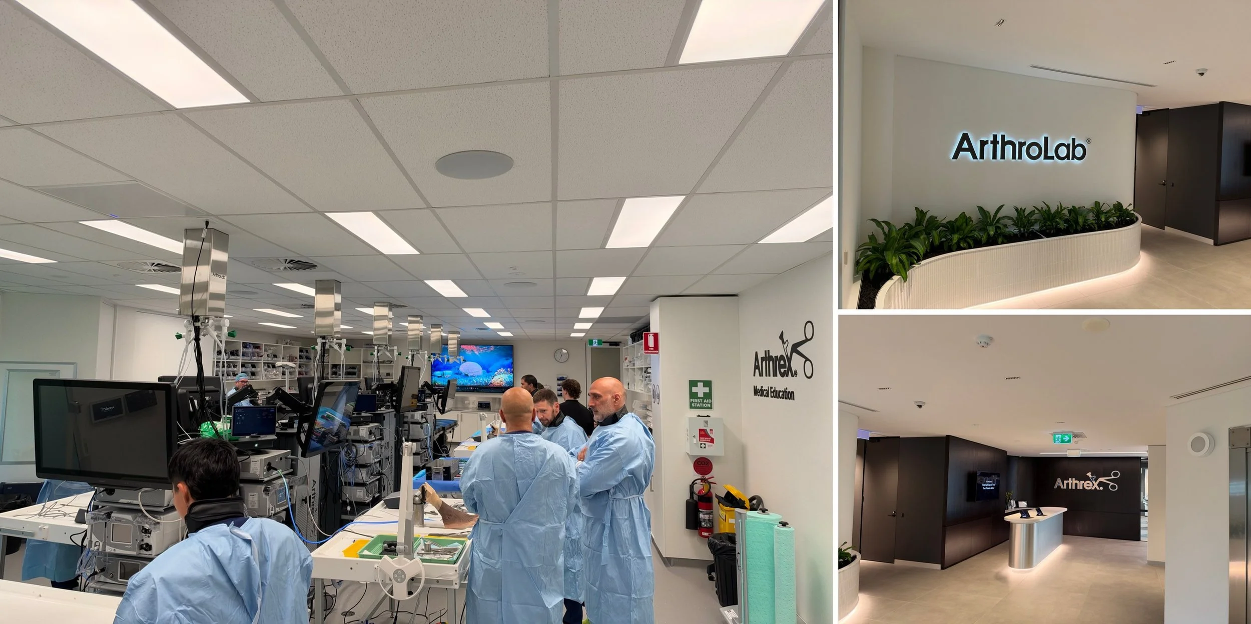
24 October: Teaching the MIS Around the Hindfoot Course
The 'MIS around the Hindfoot' course was designed for practising orthopaedic surgeons, particularly consultants in the early years of their careers who want to build confidence with minimally invasive ankle and hindfoot surgery. Dr Talia co-led the day with Sydney foot and ankle surgeon Dr Steven Kent. Short, focused lectures in the morning were followed by more than four hours of hands-on lab work.
Lateral Ankle Instability: Open or Arthroscopic Repair?
Dr Talia opened the programme with a talk on patient selection for lateral ankle ligament surgery. Chronic ankle instability after repeated ankle sprains is one of the most common problems seen in a sports-focused foot and ankle practice. Surgeons can now choose between a traditional open Broström repair and an arthroscopic (all-inside) repair.
The talk covered how to decide which patients suit each approach, what the current evidence shows when the two are compared, and how factors such as ligament quality, body weight, sporting demands and associated injuries inside the ankle joint shape that decision.
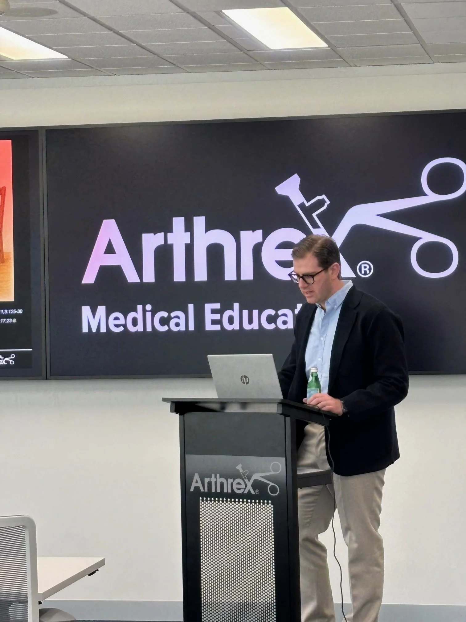
Arthroscopic Broström Repair: Technical Pearls and Augmentation
Dr Talia's second talk focused on the practical detail of arthroscopic lateral ligament reconstruction: portal placement, anchor position, ligament tensioning and lessons from his own learning curve. A key discussion point was augmentation, where a strong suture tape is added to reinforce the repaired ligament. This can be valuable for patients with poor-quality tissue, high-demand athletes and revision cases, and may support an earlier, more confident return to sport.
Syndesmosis Injuries and the Deltoid Ligament
The third talk addressed syndesmosis injuries (high ankle sprains) and the deltoid ligament on the inner side of the ankle, two areas central to Dr Talia's clinical and research work. Topics included:
• When to suspect deltoid ligament injury alongside a syndesmosis injury or ankle fracture
• What the evidence says about repairing the deltoid compared with leaving it to heal
• How to test ankle stability during surgery
• Thresholds for repair, and how these procedures can be performed through small incisions
Dr Kent added talks on minimally invasive joint preparation for ankle and subtalar fusion, correcting deformity through small incisions, and augmentation of the anterior inferior tibiofibular ligament (AiTFL), a key stabiliser of the syndesmosis.
Hands-On Cadaveric Surgical Training
Most of the day was spent in the lab, with participants rotating between two stations:
• X-ray guided MIS joint preparation, deformity correction and hindfoot fusion
• Arthroscopic and open ligament stabilisation, covering the lateral and medial (deltoid) ligaments, the syndesmosis and AiTFL augmentation
Working directly alongside faculty, surgeons practised each step, troubleshot technique and discussed real-world cases in a relaxed, collaborative setting.
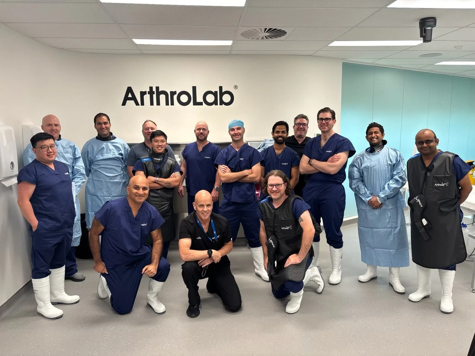
31 October: A Single-Day Lab with the Theatre Team
A week later, Dr Talia returned to ArthroLab™ for a single-day lab focused entirely on the minimally invasive procedures. These included:
• Arthroscopic lateral ankle ligament reconstruction for chronic ankle instability
• Arthroscopic and minimally invasive medial (deltoid) ligament reconstruction
• MIS hindfoot fusions, including subtalar fusion
• Minimally invasive triple arthrodesis for advanced hindfoot arthritis and deformity
Why Train as a Team?
Minimally invasive surgery depends on far more than the surgeon's hands. Each procedure involves specialised equipment: arthroscopy towers, high-speed burrs, cannulated screws, suture anchors and X-ray positioning.
Training together outside the operating theatre allows the surgeon and the team to rehearse each procedure from start to finish, standardise set-up and instrument trays, and agree on how each step will run. When the team already knows the sequence, operations can be more efficient, with less time under anaesthetic and a smoother experience for the patient.
Studies in surgical teamwork consistently show that familiar, well-rehearsed teams communicate better and work more efficiently, and that benefit ultimately flows to the patient.
This day was an investment in that teamwork. The techniques practised in Sydney are the same ones Dr Talia uses during operating lists in Melbourne.
Minimally Invasive Hindfoot Fusion and Triple Arthrodesis
For patients with arthritis or severe deformity of the hindfoot, a fusion (arthrodesis) can provide lasting pain relief and a stable, plantigrade foot. A triple arthrodesis fuses three joints of the hindfoot: the subtalar, talonavicular and calcaneocuboid joints. Traditionally this needed large incisions, which carry a risk of wound-healing problems, particularly on the outer side of the foot.
Using MIS techniques, the joint surfaces can be prepared through small portals with a burr under X-ray guidance, and the fusion held with screws placed through small incisions. The aim is to achieve a solid fusion while protecting the skin and soft tissues and reducing wound complications.
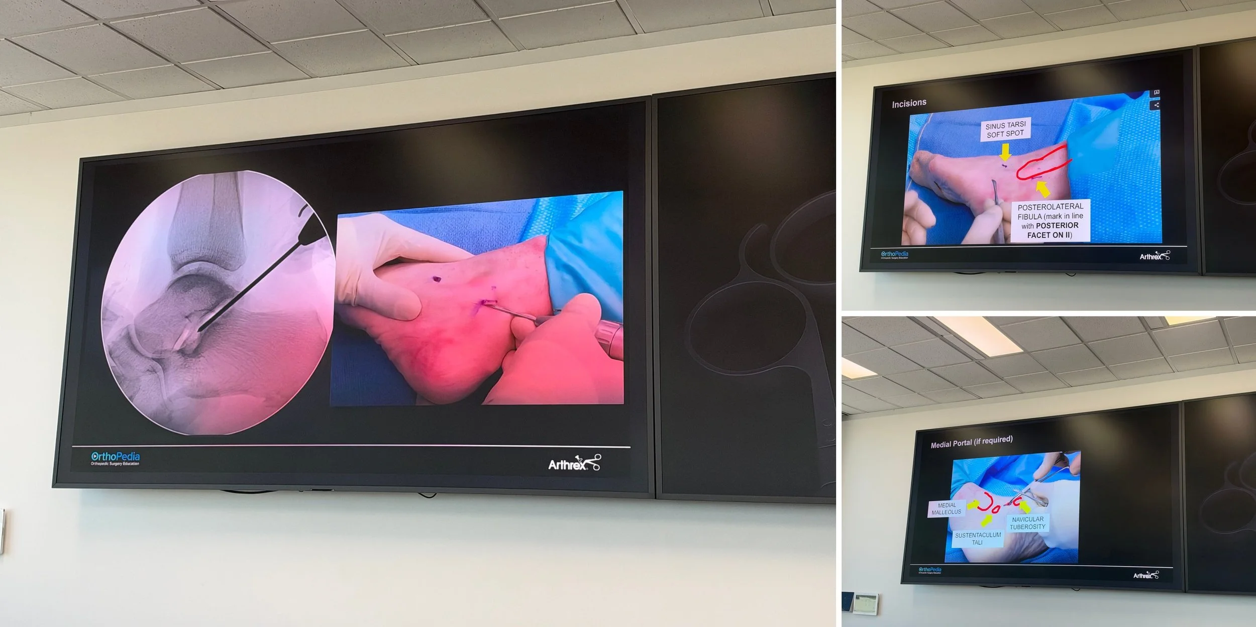
Who May Benefit from Arthroscopic Ankle Ligament Reconstruction?
Most ankle sprains settle with physiotherapy and a structured rehabilitation programme. Surgery is generally considered when the ankle continues to give way despite good rehabilitation, or when instability is limiting sport, work or day-to-day activity.
Arthroscopic reconstruction has the added advantage of allowing the surgeon to inspect the inside of the ankle joint at the same time. Cartilage injuries, scar tissue, bony spurs and loose bodies are common in unstable ankles and can often be treated during the same operation. Medial (deltoid) ligament injuries can also be identified and addressed, which is important because combined medial and lateral instability is easily missed.
Recovery After Minimally Invasive Foot and Ankle Surgery
Recovery depends on the procedure performed. After arthroscopic ankle ligament reconstruction, most patients spend a short period in a boot before starting a guided physiotherapy programme focused on range of motion, strength and balance. Return to running and sport is gradual and tailored to each person.
Hindfoot fusions and triple arthrodesis require a longer period of protection while the bones heal. Smaller incisions do not shorten the time it takes for bone to fuse, but they may reduce pain, swelling and wound complications in the early weeks. Every patient receives an individual recovery plan, and Dr Talia works closely with physiotherapists and the patient's GP throughout.
What This Means for Patients
Not every patient is suited to minimally invasive surgery. Severe instability, poor ligament tissue, significant deformity or advanced arthritis may still be best treated with an open approach. The goal is always to choose the right operation for each person, based on their condition, goals and the best available evidence.
What patients can expect is a surgeon trained in the full range of options, from arthroscopic ankle ligament reconstruction to minimally invasive hindfoot fusion, supported by a theatre team trained in the same techniques.
Dr Adrian Talia's Commitment to Orthopaedic Education
Dr Talia is a fellowship-trained foot and ankle surgeon with international training from Oxford and Basel. Alongside his private practice at Melbourne Orthopaedic Group in Windsor and his clinical appointment at Western Health, he is actively involved in research, registrar teaching and surgical skills courses across Australia and internationally.
Dr Talia thanks Arthrex Australia, Rick Osler and the Arthrex Medical Education team, Dr Steven Kent, all participating surgeons for two excellent days at ArthroLab™.
If you have ongoing ankle instability, a syndesmosis or deltoid ligament injury, or hindfoot arthritis, contact Dr Talia's rooms on (03) 9124 7960 or talia.admin@mog.com.au.